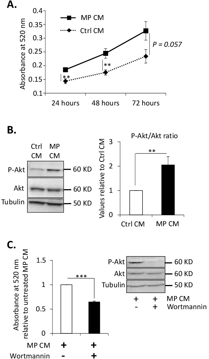
Fig. 1. The MP-dependent stimulation of FAP proliferation is mediated by Akt signaling. FAPs proliferated in the presence of conditioned medium (CM) from MPs (MP CM) compared to control conditioned medium (Ctrl CM). (A) Proliferation of FAPs was measured by MTT assays 24 hours, 48 hours and 72 hours after plating (n=5 donors). (B) Confluent FAPs were treated with Ctrl CM or MP CM and total proteins were extracted 15 minutes later. Protein expression of phosphorylated Akt (P-Akt), total Akt (Akt) and tubulin was assessed by western blot (left panel). P-Akt band intensity was quantified and normalized to total Akt signals (right panel) (n=3 donors). (C) FAPs proliferated in the presence of MP CM with or without wortmannin. Proliferation of FAPs was measured by MTT assays 48 hours after plating. Protein levels of P-Akt, total Akt and tubulin was assessed by western-blot. Representative immunoblot is shown (n=3 donors). *** P<0.001; ** P<0.01; p values close to significance are indicated.